Fracture Analysis of Concrete Using Scanning Electron Microscopy
Fracture Analysis of Concrete Using Scanning Electron Microscopy
Kamran M. Nemati
Postdoctoral Research Fellow
Department of Civil and Environmental Engineering
University of California at Berkeley
Berkeley, California 94720 U.S.A.
Summary:
A special experimental technique has been developed which makes possible the preservation of the compressive stress-induced microcracks in concrete as they exist under applied loads. This technique involves injecting a molten-metal alloy into the induced cracks and solidifying it before unloading. Scanning electron microscopy (SEM) was employed to capture images from the cross sections of the concrete specimens. These images were then used to study the generation and interaction of the compressive stress-induced microcracks and the effect of confinement on microcrack behavior.Keywords:
Concrete, Stress-induced microcracks, Scanning Electron Microscope (SEM), image analysis, Wood's metal.Introduction
The scanning electron microscope (SEM) is one of the most versatile instruments available for the examination and analysis of microstructural characteristics of solid objects. The primary reason for the SEM's usefulness is the high resolution that can be obtained when bulk objects are examined.
The microscope has been a powerful tool in the study of cement and concrete since the early development of these materials. Le Chattelier (1882) was amongst the first to apply the microscope to the study of cementitious materials. He used it to investigate the chemical and physical aspects of hydration and setting, rather than to study cracks. His efforts undoubtedly influenced later workers in their use of microscope. Tavasci (1942) successfully used the microscope to study the composition and structure of concrete, but not for cracks per se. His work, however, set the stage for the studies of cracks on the interior surfaces of cut specimens which were conducted in the 1960s.
The electron microscope was apparently first used by Eitel (1941, 1942), and by Radczewski and co-workers (1939) to study the hydration process of concrete. Grudemo (1960) was another important pioneer in the use of high magnification, including the use of electron microscope. Although most of these studies were not directly related to cracks, the led the way to later studies of cracks in which electron microscopy was a powerful tool. Diamond and Mindess (1980) used the scanning electron microscope to observe the growth of surface cracks during loading, using magnifications generally from 35
x to 450x.This investigation of microcracking ranges from a macroscopic studies of the behavior of cracked specimens to a microscopic study of the cracks themselves. The presence of microcracks was predicted on the basis of macrobehavior and verified by microscopic study. Several methods have been used to study the microcracking of concrete. Some of those methods are acoustic emission, sonic testing, microscope and
x-ray techniques, hydrophilic tracer liquid technique, dye technique, lead salt, microscope technique with dye, mercury intrusion porosimetry, x-ray technique, computerized tomography analysis, fiber optics, and holographic interferometry. Almost all of the above-mentioned techniques failed to render accurate representations of the geometry and state of microcracks as they exist under load. Some of the techniques are limited in their resolution, sensitivity in detecting cracks, and ability to make full-field observations. Other methods are incapable of examining the specimen while under load; or they require special preparation of the specimen, which alters its behavior.Materials and Methods
The method utilized here involves the application of a metal in liquid phase, Wood's metal, which has a melting point range from 70° C to 88° C to preserve the microstructure of stress-induced microcracks in concrete. Wood's metal has a Young's modulus of 9.7 GPa, a density of 9.4 g/cm3, and an effective surface tension of about 400 mN/m (Yadev et al. 1984), and is solid at room temperature. The advantage of such an alloy is that it can be injected into voids and stress-induced microcracks at the desired stress level, then solidified to preserve, in three-dimensional form, the geometry of the microcracks induced at any given stage of the experiment. Used in conjunction with scanning electron microscopy, it has made possible the detailed observation of microcracks in concrete as they exist under load. Named after the astronomer who used an alloy of bismuth, lead, tin, and cadmium to create a perfect parabolic surface for astronomical observations, Wood's metal has been used in the past few years to study the microstructure of different materials (Yadev et al. 1984, Pyrak.1988, Zheng, 1989, Nemati 1994, Chang et al. 1996).
The objectives of this investigation is to examine the mechanisms for the generation of stress-induced microcracks in concrete under compression, and establish the effect of microstructure on the generation of stress-induced microcracks, to determine the real shapes and geometry of stress-induced microcracks as they exist under load. This was accomplished by injecting a molten metal into the induced cracks and solidifying it before unloading. The test equipment created to preserve the cracks under applied load is described in detail elsewhere (Nemati 1994). Cylindrical concrete specimens were tested in compression under uniaxial loading and with lateral confinement. A no-load experiment was conducted initially to determine the fracture status of the specimen prior to loading. In the ensuing experiments, the specimen was subjected to uniaxial and triaxial compression. Confinement was used to generate triaxial compression and was supplied by stainless steel wires wound around the concrete specimens. While under load, the specimens were impregnated with Wood's metal to preserve the induced cracks. In order to saturate the induced microcracks with the molten metal, a nitrogen pressure of 10.3 MPa was applied to the molten metal as the pore pressure, which was kept constant throughout the tests. After the metal solidified, sections were cut from the cylinders.
The next step was to polish the specimens to be examined in a scanning electron microscope. The specimens extracted from the concrete cylinders were 25 mm square and had an approximate thickness of 5 mm. First, one side of each specimen was polished with 120#, 220#, 320#, and 600# silicon carbide using a rotating grinder and mounting it against a 25 mm-diameter glass plate with epoxy. In order to make both sides of the specimen parallel to each other, the samples were cut 2-3 mm thick by using a diamond slicing wheel with a nonaqueous lubricant (Propylene glycol coolant). The specimen were then lapped with a wheel grinder and polished with 600# silicon carbide. Further polishing was performed with 100-, 50-, and 10-micron aluminum powder on a glass plate. The final stage involved was treating specimens with 5-, 3-, and 1/4-micron diamond paste using a special polishing equipment. After each stage of polishing, the specimens were immersed in acetone and placed in an ultrasonic bath in order to remove the residual silica film on their surfaces, thus preparing them for the next stage of polishing. The specimens were studied using a jeol jsm-35cf scanning electron microscopy (SEM) in conjunction with a kontron SEM-ips image analyzer. Image analysis was used to characterize the quantity and distribution of cracks in concrete under various loading conditions.
Results
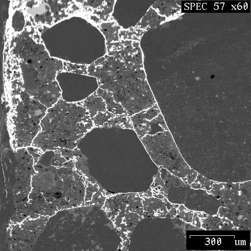
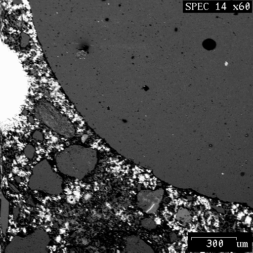
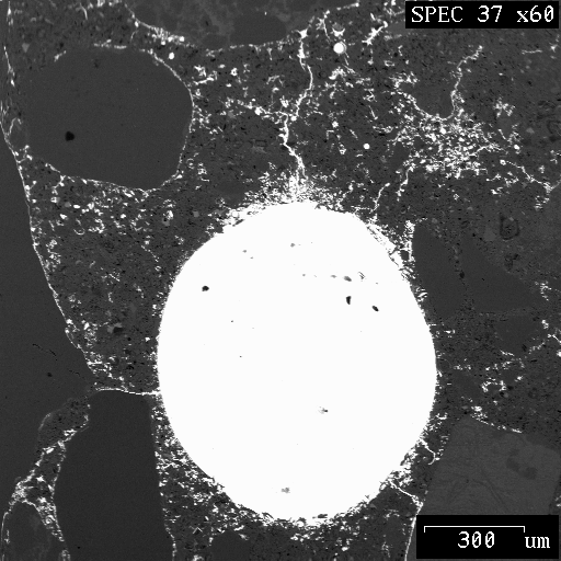
After the concrete samples were prepared for scanning electron microscope studies, images were extracted from each sample. A total of four samples taken from the center and edge of the concrete cylinders in axial direction were studied. The images were acquired by the image analyzer at a magnification of ´ 60 and digitized into an array of 512 ´ 512 pixels with 256 gray levels (1 pixel = 3.3 µm). A typical gray level backscattered electron (BSE) image is shown in Figure 1.
|
|
|
|
Figure 1 A typical BSE image |
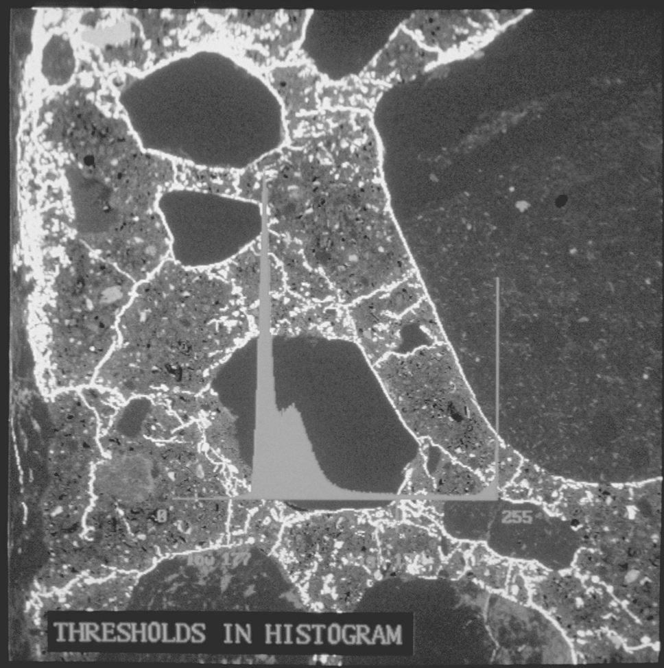
Figure 2 Establishing threshold in histogram |
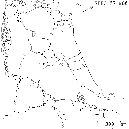
Figure 2 shows a histogram of the distribution of gray levels in the BSE image, superimposed on the original image. The peak at the right (high gray level) corresponds to the areas of Wood's metal, while the peaks to the left correspond to the cementitious phases and aggregate. This histogram was used to select the threshold values for discriminating the areas of Wood's metal. Figure 3 is the final binary image used for crack analysis.
|
|
|
Figure 3 Crack network |
Figures 4 shows typical images from the unloaded specimen. In order to restrain the specimen during the impregnation with Wood's metal this specimen was subject to a small load of 10 MPa. In this specimen the bright areas which have been permeated by Wood's metal correspond to regions of connected pores or microcracks. It is important to note that concrete is inherently cracked even before the application of any load (Hsu et al. 1963). Existing pre-load cracks are due to factors such as bleeding, creep, drying shrinkage, etc. However, there are no long cracks characteristic of stress-induced cracking.
|
|
|
|
Figure 4 SEM micrographs from the no-load specimen |
|
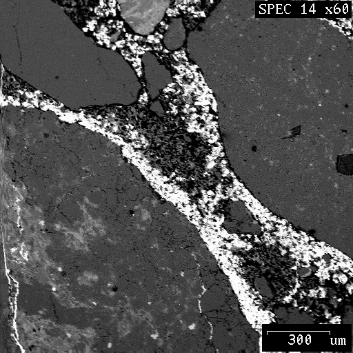
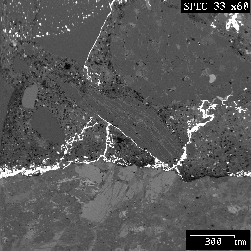
The microcracks in the rest of the specimens appear to have been generated by several different mechanisms. Figure 5 shows two micrographs taken from a specimen under laterally confined condition. As can be seen, microcracks propagate through the cement paste and along the transition zone, which is the interface between the aggregate and the cement paste.
|
|
|
|
Figure 5 SEM micrographs from the laterally confined specimen |
|
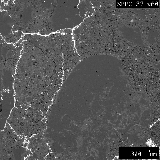
Many cracks were generated from voids. Figures 6 shows a micrograph of microcracks generated from a pore space, which is filled with Wood's metal. These microcracks were generated as a result of local tensile stress tangential to the boundary of voids, with a value that was of the order of the maximum applied principal stress. It was found that these cracks usually start from the pore boundaries and then propagate in the direction of maximum compression.
|
|
|
|
Figure 6 SEM micrographs of microcracks propagating from a pore |
|
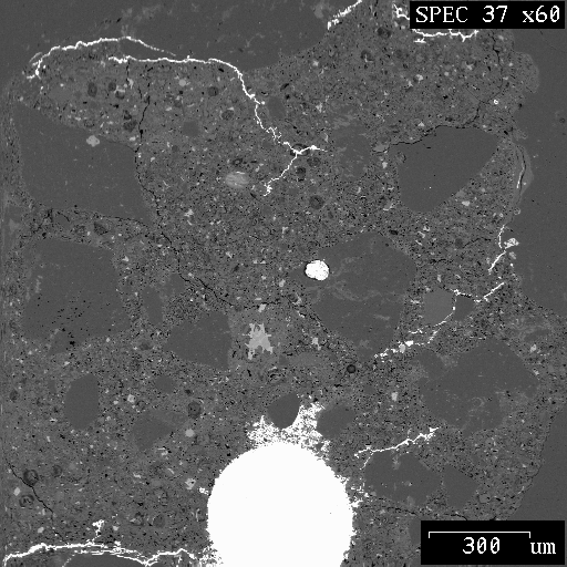
Microcracks were also found to have been generated from the inside of aggregates, in a manner similar to a Brazilian test, when the aggregates were loaded across their height. Figure 7 shows a micrograph of this phenomenon.
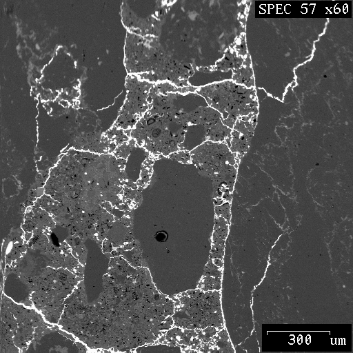
Figure 7 SEM micrograph of aggregate cracking
By submerging the specimens in acid, the network of fractures in concrete can be observed in three-dimensional form as shown in Figure 8.

Figure 8 SEM micrograph of the network of fractures in three-dimensional form
Discussion
A special experimental technique was developed which made possible the preservation of the compressive stress-induced microcracks in concrete as they exist under applied loads. This technique involved injecting a molten-metal alloy into the induced cracks and solidifying it before unloading.
The use of scanning electron microscopy (SEM) to extract images from the concrete specimens for different testing conditions is explained. The images rendered were analyzed using an image analyzer. The image analyzer identifies Wood's metal, which represents the crack network in concrete specimens. The scanning electron microscope identifies the geometrical aspects of features in the microstructure, such as microcracks in concrete represented by Wood's metal.
References
Chang, C.T., Monteiro, P., Nemati, K., Shyu, K. (1996). "Behavior of Marble Under Compression", Journal of Materials in Civil Engineering, Vol. 8, No. 3, 157-170.
Diamond, S., and Mindess, S. (1980) "A Preliminary SEM Study of Crack Propagation in Mortar", Cement and Concrete Research, Vol. 10, No. 4, 509-519.
Eitel, W. (1941). "Recent Results of Cement Research," Angewandte Chemie, Vol. 54, No. 15/16, 185-192.
Eitel, W. (1942). "Electron Microscopic Cement Research," Zement, 31, 489-495.
Grudemo, A. (1960). "The Microstructure of Hydrated cement Paste," Proceedings, Fourth International Symposium on the Chemistry of Cement, 2, 615-658, Washington, D.C.
Hsu, T. T. C.; Slate, F. O.; Sturman, G. M.; and Winter, G. (1963). "Microcracking of Plain Concrete and the Shape of the Stress-Strain Curve," Journal of the American Concrete Institute, Proceedings, 209-224.
Le Chatelier, H. (1882). "Experimental Researches on the Constitution of Hydraulic Mortars," translated by J. L. Mack, McGraw, New York, 1905. Also: Compt. rend., 94 (1882) 13; and Journal of Chemical Ind., 1 (1882) 151.
Nemati, K.M. (1994). "Generation and Interaction of Compressive Stress-Induced Microcracks in Concrete," Ph.D. Thesis, Department of Civil Engineering, University of California at Berkeley.
Pyrak, L. J. (1988). "Seismic Visibility of Fractures." Ph.D. Thesis, Department of Material Science and Mineral Engineering, University of California at Berkeley.
Radczewski, O. E.; Müller, H. O.; and Eitel, W. (1939). "Ultra-Microscopic investigation of the Hydration of Free Lime," Zement, Vol. 28, No. 49, 693-697.
Tavasci, B. (1942). "Structure of Hydrated Portland Cement," Zement, Vol. 30, No.4, 43-48 & 55-58.
Yadev, G.D., Dullien, F. A. L., Chatzis, I., and Macdonald, I. F. (1984). "Microscopic Distribution of Wetting and Non-Wetting phases in Sandstone During Immiscible Displacements," Paper SPE 13212, presented at the 1984 SPE Annual Technical Conference and Exhibition, Dallas, Texas.
Zheng, Z. (1989). "Compressive stress-induced microcracks in rocks and applications to seismic anisotropy and borehole stability," PhD Thesis, Department of Material Science and Mineral Engineering, University of California at Berkeley.
Address for reprints:
Kamran M. Nemati, Ph.D.